Sacral OCD
Case example
An obstacle in the tunnel
A 3 year-old, female mixed breed dog was presented for pelvic limb lameness of six months duration.
CT findings
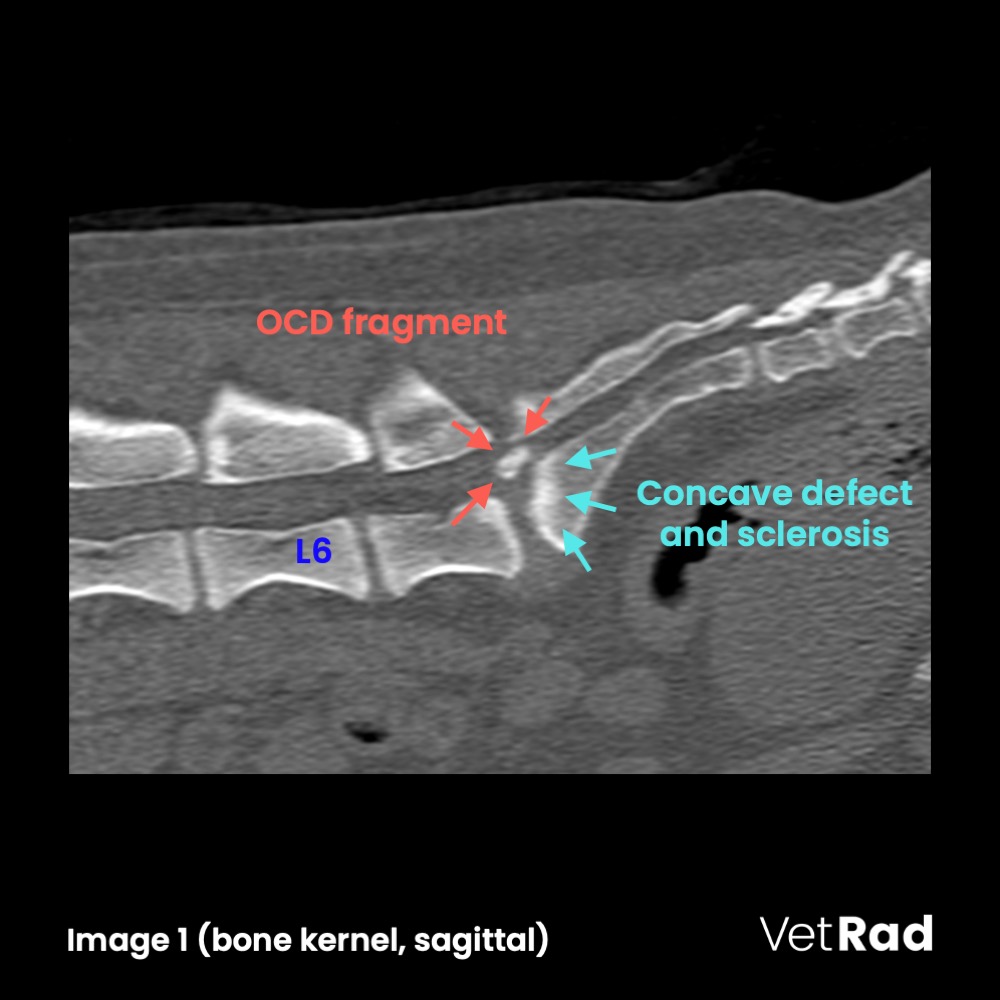
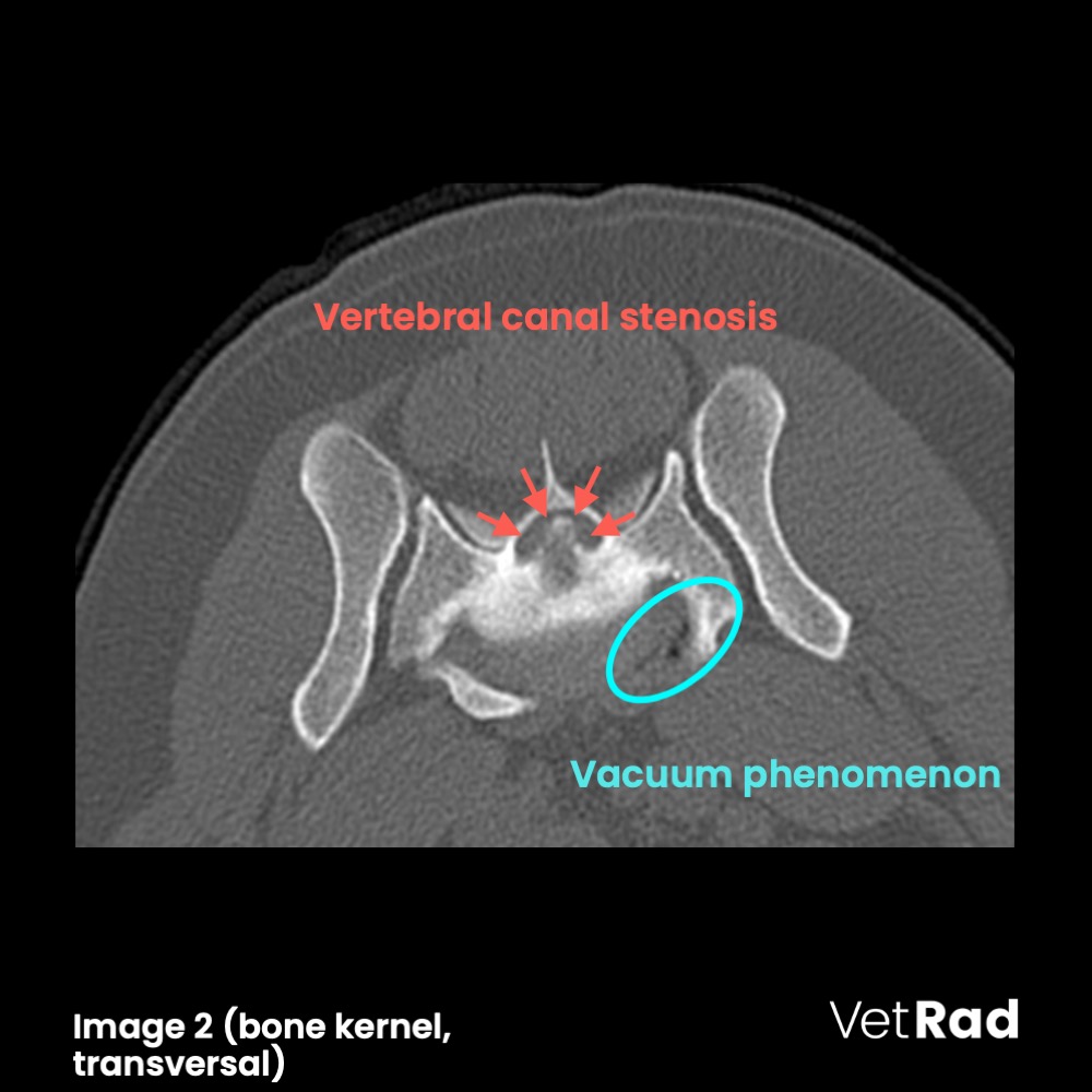
The craniodorsal contour of the cranial sacral extremity is blunted with moderate, adjacent sclerosis. An oval, smoothly contoured, heterogeneous mineral attenuation (image 1) is craniodorsally adjacent, occupying the near entirety of the vertebral canal, causing dorsal deviation and near complete indistinction of the cauda equina at this level.(image 2) The intervertebral disc/annulus fibrosus is enlarged and surrounds the aforementioned mineral attenuation contributing to the vertebral canal stenosis. A vacuum phenomenon is within the left ventral aspect of the narrowed lumbosacral intervertebral disc.(image 2) The caudal vertebral endplate of L7 is moderately sclerotic and lumbosacral spondylosis deformans is evident.
Conclusions
- Sacral osteochondritis dissecans, causing severe, extradural lumbosacral stenosis.
Take Home
- Radiography is most often diagnostic, but cross-sectional imaging is of higher diagnostic value as it allows for a more precise localization of the osteochondral fragment, as well as a more detailed evaluation of the degree of vertebral canal and/or intervertebral foraminal stenosis and scrutinization for involvement of the cauda equina/segmental nerves.
- Vacuum phenomena characterize aseptic gas collections of nitrogen and a small portion of oxygen or carbon dioxide, classified as intradiscal, intraosseous and intraarticular. They can be seen in normal or pathologic tissues in the scope of aging or in areas of distractive forces.
More information
- »Sacral osteochondrosis in two German Shepherd Dogs«
— Mathis et al., Aust. Vet J. 2019 Jan;10:169-183 - »Lumbosacral osteochondrosis: radiological features and surgical management in 34 cases«
— Hanna, JSAP 42(6) 2001; 272-278 - »Vacuum Phenomenon of the Canine Spine: CT findings in 3 patients «
— Hathcock, Vet Radiol and Ultrasound 1994; 35(4); 285-289
Images courtesy of the AniCura Tierklinik Hollabrunn, Austria.
UPLOAD MEDICAL IMAGES NOW