Trigeminal Nerve Neoplasia
Case example
Nerve dumbbells do not make one stronger
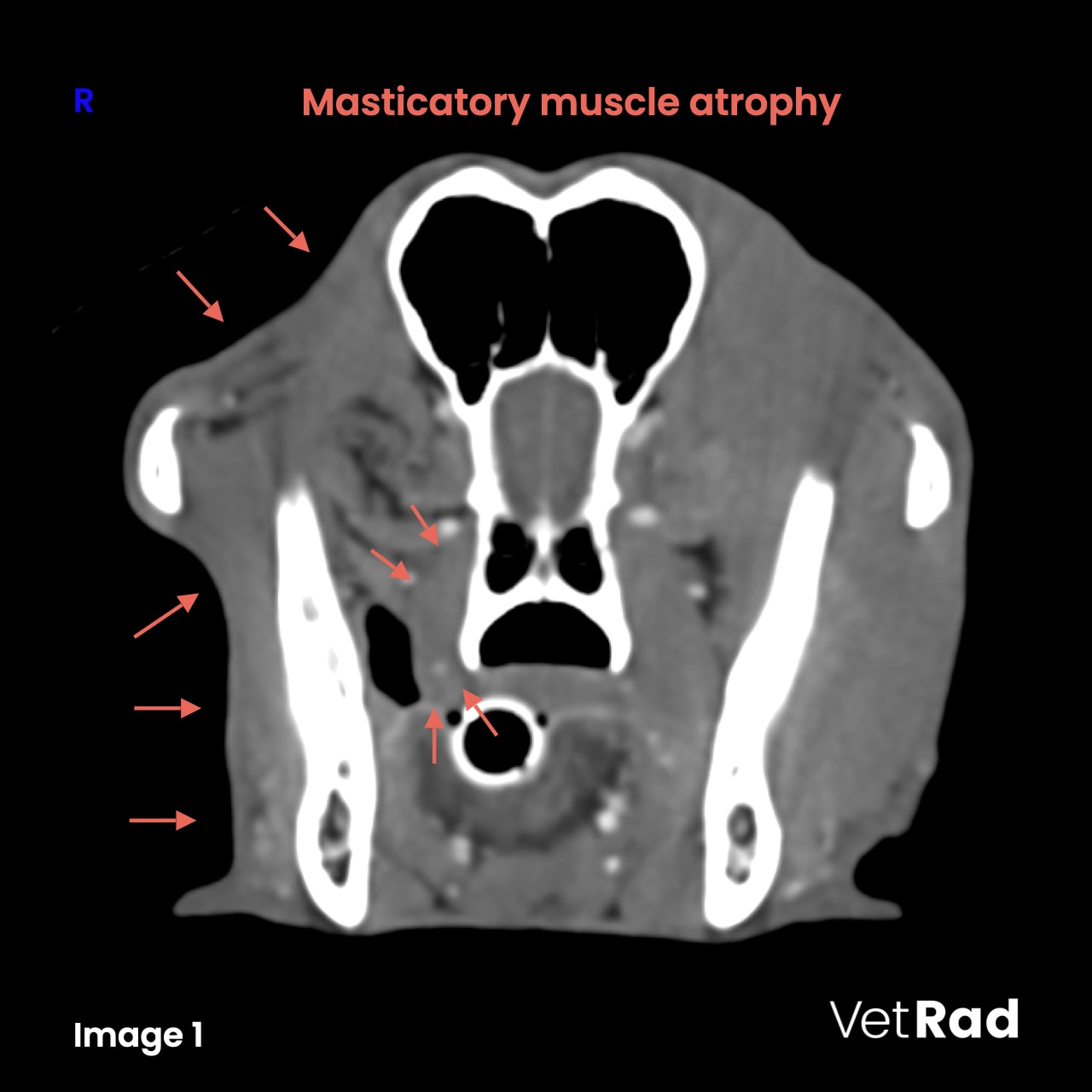
A 10 year-old, male intact mixed breed dog was presented with acute right sided muscular atrophy of the head and facial paresis.
CT findings
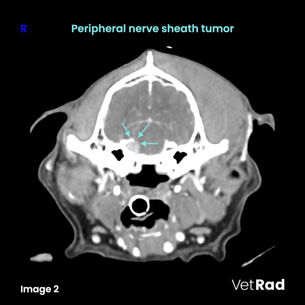
A moderate reduction in size of all right masticatory muscles (M. temporalis, M. masseter, M. pterygoideus medialis et lateralis and M. digastricus) is evident. (Image 1) The right ocular bulb is retracted. A moderately enlarged, tubular and mildly heterogenously contrast enhancing region is seen along the course of the trigeminal nerve canal.(Image 2) The oval foramen is ipsilaterally mildly enlarged.


Conclusions
- Peripheral nerve sheath tumor (PSNT) of the right trigeminal nerve with secondary ipsilateral masticatory muscle atrophy. Differential diagnoses include a schwannoma or neurofibroma.
Learning points
- Although magnetic resonance imaging (MRI) is the modality of choice for diagnosing cranial nerve neoplasia, computed tomography (CT) can be a useful alternative.
- PNST are presented as an extra-axial mass with or without distortion of the associated brainstem, while trigeminal nerve neuritis is usually characterized by diffuse and less pronounced enlargement. Presence of fluid in the ipsilateral tympanic bulla, due to dysfunction of the auditory tube is also more common in trigeminal nerve neoplasia.
More information
- »Clinical features of trigeminal nerve-sheath tumor in 10 dogs«
— J Am Anim Hosp Assoc 1998;34(1):19-25. - »Clinical and imaging findings, treatments, and outcomes in 27 dogs with imaging diagnosed trigeminal nerve sheath tumors: A multi-center study«
— Vet Radiol Ultrasound 2017;58:679-689.
Images courtesy of the Veterinary Clinic Tulln, Austria.
UPLOAD MEDICAL IMAGES NOW

