Craniomandibular osteopathy
18 May, 2021
Case example
Cheeky cheek
A 10-month-old Newfoundland dog presents with a thickened left mandible.
CT findings
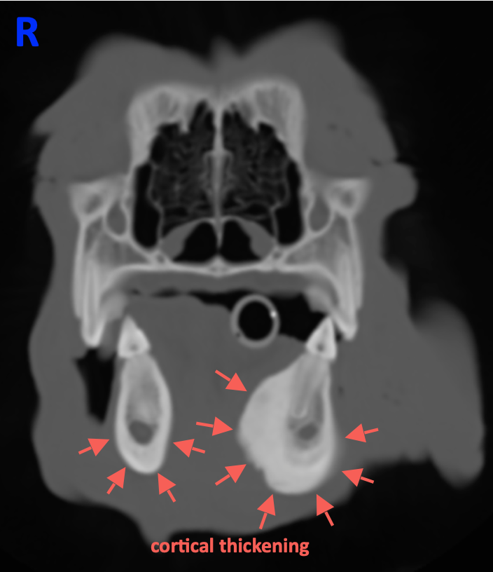
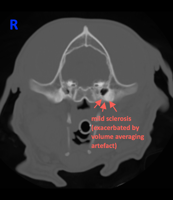
The left mandible shows moderate thickening of the cortex with smooth, broad-based periosteal proliferations (Image 1). The cortex of the contralateral side is also mildly thickened (Image 1). The tympanic bulla and the region of the Proc. retroarticularis are sclerotic bilaterally (Image 2).


Conclusions
- Bilateral, asymmetric craniomandibular osteopathy (CMO)
Learning points
- A thickened cortex of the mandible and/or the tympanic bullae in young dogs are typical signs of CMO
- Radiographic appearance can confirm the suspected clinical diagnosis
- West Highland White Terrier is overrepresented; other breeds are also described
More information
- »Hudson JA, Montgomery RD, Hathcock JT, et al. Computed tomography of craniomandibular osteopathy in a dog«
— Vet Radiol Ultrasound 1994;35:94–99. - »Craniomandibular osteopathy in two Pyrenean mountain dogs«
— Vet Rec 1998;142(17):455-9.
Images courtesy of the Anicura Tierklinik Hollabrunn, Austria.
MEINE DATEIEN JETZT HOCHLADEN

