Insulinoma
25 September, 2019
Case example
When the pancreas glows briefly
A 6-year-old, male mixed breed dog was presented for progressive lethargy and a recent syncope. The most significant blood work finding was hypoglycemia.
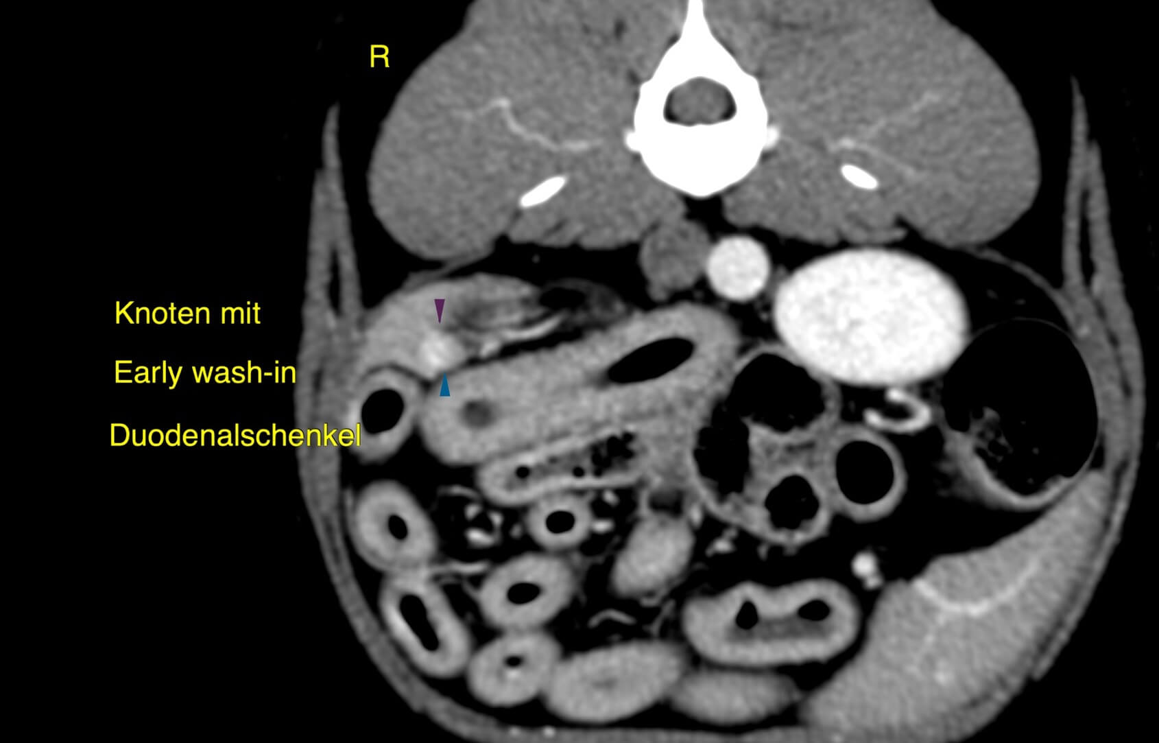
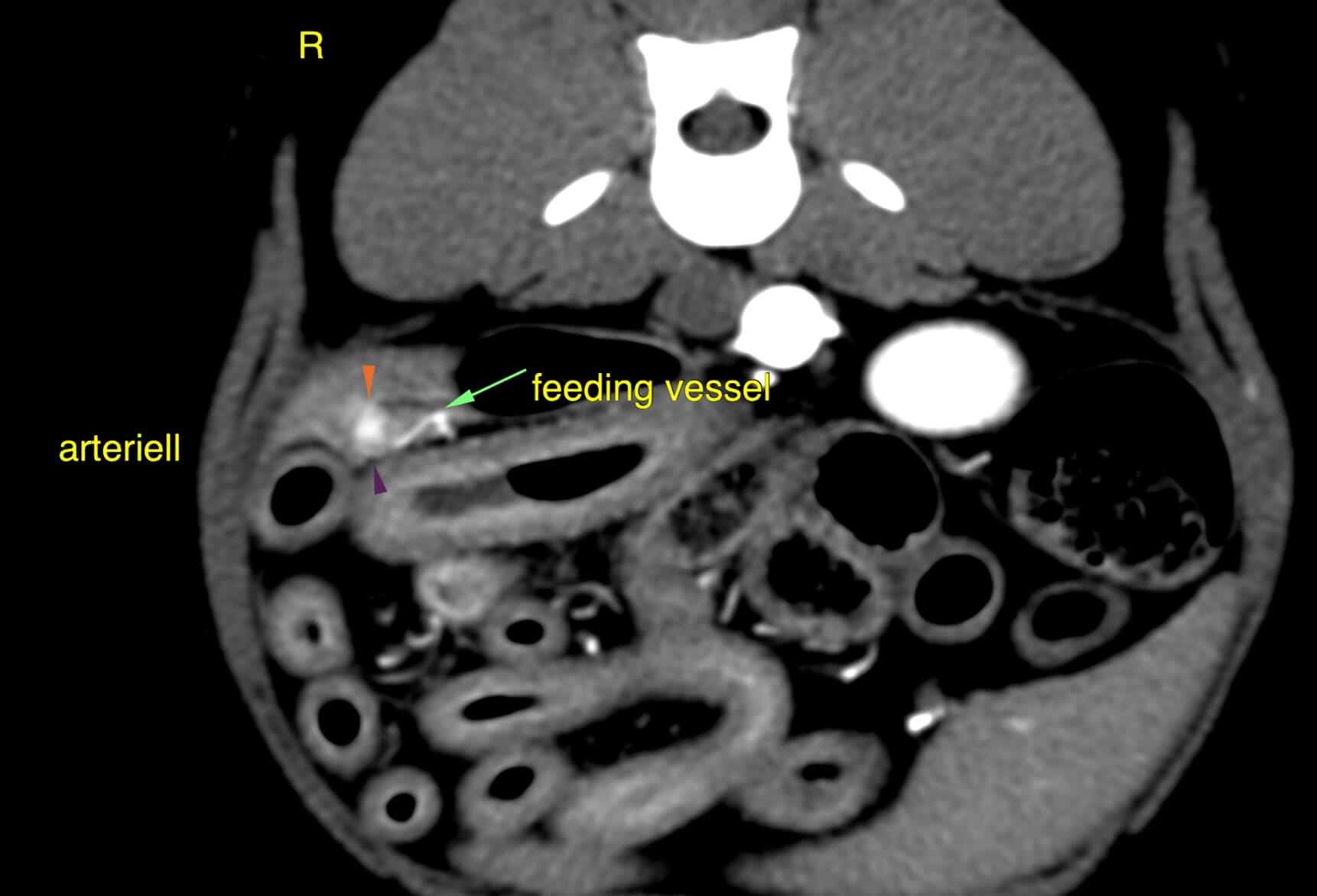
CT findings
A small 6 mm nodule is seen within the caudal aspect of the right pancreatic lobe with evidence of an „early-wash-in and early-wash-out” enhancement pattern. This nodule is most conspicuous in the arterial phase after intravenous contrast administration and subsequently shows the same enhancement characteristics as the remaining pancreatic parenchyma. A small tortuous vessel is evident entering the pancreas from the periphery, likely representing a tumor feeding vessel.
Conclusions
Given the clinical presentation and laboratory workup results, an insulinoma (canine pancreatic neuroendocrine tumor) is the most likely differential diagnosis.
Learning points
- Given the small size and poor distinction form the surrounding pancreatic tissue, CT- Angiography has been the most valuable diagnostic imaging tool in diagnosing canine pancreatic neuroendorine tumors. In addition, insulinomas are hypervascular carcinomas deriving from the pancreatic islet cells with marked enhancement during the arterial phase, contrasting the surrounding, mildly enhancing pancreatic tissue.
- Delayed imaging may render insulinomas invisible against the surrounding tissue
- It is of outmost importance to catch an early phase of perfusion whenever insulinoma is a potential
Literature
- “Utility of contrast-enhanced computed tomography in the evaluation of canine insulinoma location” Vet Q. 2018 Dec; 38(1):53-62
- “Characterization of triple-phase computed tomography in dogs with pancreatic insulinoma” J Vet Med Sci. 2016;77(12):1549-53
- “Dynamic Computed Tomography of the pancreas in normal dogs and in a dog with pancreatic insulinoma” Vet Radiol Ultrasound 2007 Jul-Aug;48(4):328-31
Images courtesy of Tierklinik Haar
UPLOAD MEDICAL IMAGES NOW