Retrobulbar abscess
18 June, 2020
Case example
Retrobulbar abscess – When the eye protrudes
A 15-year-old, female-spayed Podenko was presented for an acute right exophthalmos and pain upon opening its mouth.
CT findings
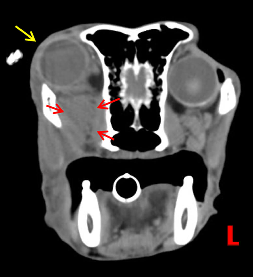
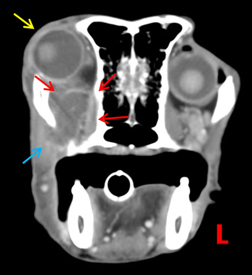
An amorphous fluid to soft tissue attenuating region is within the ventral and axial aspect of the right retrobulbar space in the area of the well-defined zygomatic gland and medial pterygoid muscle. Ring-enhancement is evident and a mild to moderate mass-effect causes rostrodorsal displacement of the ipsilateral globe (image 1 & 2).
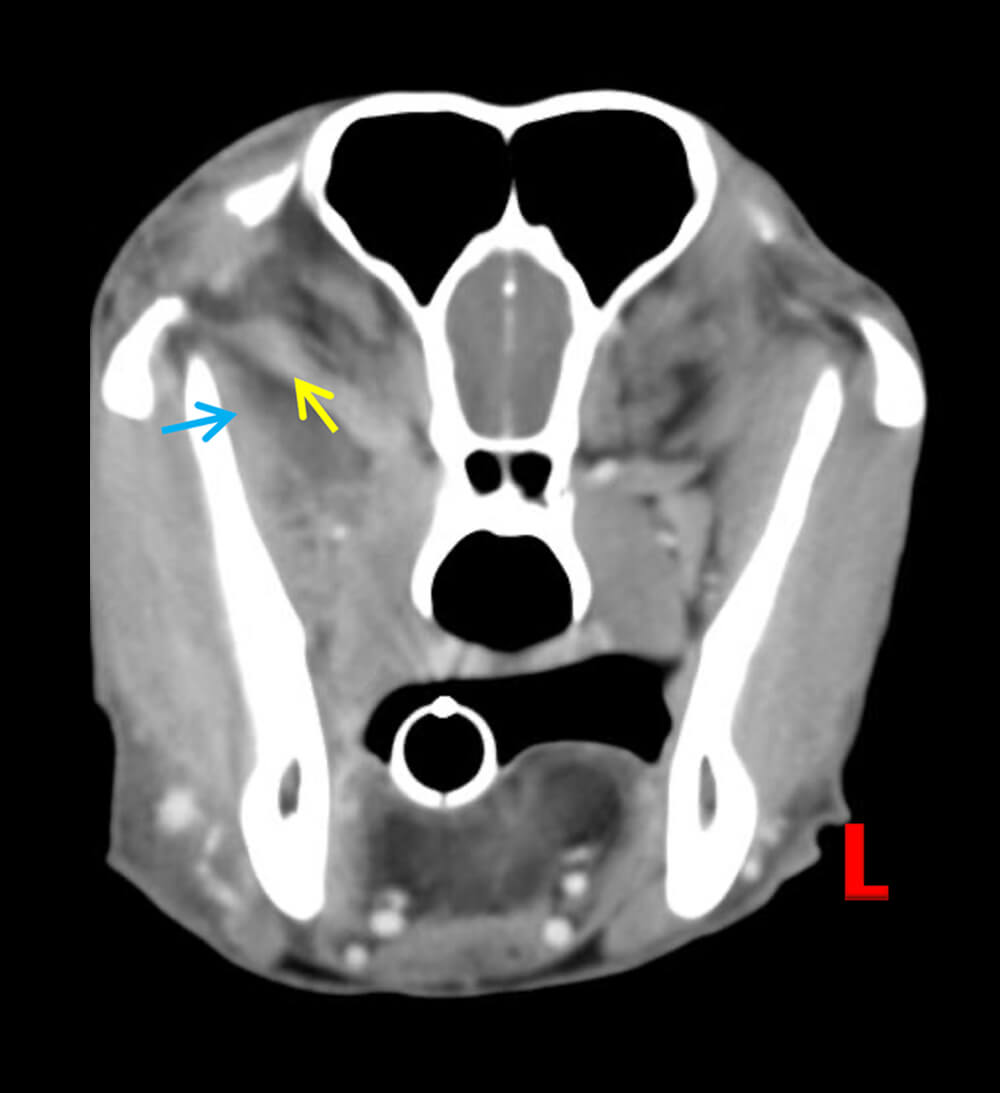
There is moderate and homogenous contrast-enhancement of the right extrinsic ocular and masseteric muscles (image 3). The right mandibular and medial retropharyngeal lymph nodes are mildly enlarged and homogenously contrast enhancing.
Conclusions
The acute right exophthalmos is secondary to a retrobulbar abscess with regional myositis and mild, reactive lymphadenopathy. There is no overt evidence of a foreign body within the associated soft tissues, regional penetrating trauma, zygomatic sialadenitis or a tooth-root abscess.
Learning points
- Computed Tomographic Angiography can aid in the diagnosis of retrobulbar processes and has been most helpful in detecting wooden foreign bodies in comparison to ultrasonography and even Magnetic Resonance Imaging. Typical characteristics include a mass-effect, ring-enhancement, enhancement of the regional musculature and regional lymphadenomegaly.
- However, note that foreign bodies, such as grass-awns, wooden foreign bodies or other soft tissue attenuating foreign material may not be evident even on Computed Tomography due to the similar attenuation values to fat and/or soft tissues.
- Typical findings in malignant neoplasia include regional osseous lysis, which is not evident in this case.
- “Retrobulbar cellulitis and abscessation: focus on short- and long-term concurrent ophthalmic disease in 41 dogs” MC Fischer et al. J Small Animal Pract. 2018; Dec;59(12):763-768
- “Clinical Features and Computed Tomography Findings are Utilized to Characterize Retrobulbar Disease in Dogs” JN Winer et al., Front. Vet. Sci. August 2018
- “Ultrasonographic findings in 50 dogs with retrobulbar disease” DR Mason et al. JAAHA. 2001;37 6 557-562
Images courtesy of the Tierklinik Posthausen, Germany
UPLOAD MEDICAL IMAGES NOW