Traction bronchiectasis
31 May, 2019
Case example
A 15 year-old cat presents with chronic cough and polypnea. There was no improvement after antibiotic treatment.
CT findings
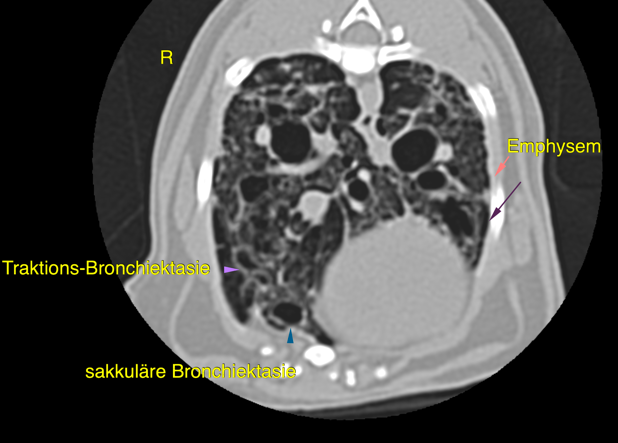
The CT of the thorax reveals severe generalized pulmonary interstitial changes with saccular bronchiectasis, traction bronchiectasis (Figure 1), multifocal honeycombing (Figure 2) and subpleural infiltrates and interstitial bands with diffuse pulmonary emphysema. (Figure 3)
Conclusions
These findings support the presence of pulmonary fibrosis and can be secondary to chronic bronchitis, such as feline lower airway disease, idiopathic pulmonary fibrosis, as well as chronic infectious causes. The prognosis is poor owing to irreversibly decreased parenchymal compliance, reduced mucociliary clearance and presumably impaired gas exchange. Secondary superinfection is a common risk and to date no curative treatment option exists.
Learning points
Traction bronchiectasis refers to pathologic dilatation of bronchi, caused by traction of surrounding parenchymal fibrosis. It is a subtype of bronchiectasis secondary to distortion of the lung parenchymal architecture. In this case it is characterized by severe structural pulmonary changes without evidence of a classic bronchial pattern.
Literature
„Further characterization of computed tomographic and clinical features for staging and prognosis of idiopathic pulmonary fibrosis in West Highland white terriers” Thierry et al., Vet Radiol Ultrasound 2017; 58: 318-388
“A case of atypical diffuse feline fibrotic lung disease” Le Boedec et al. J Feline Med Surg, 2014 Oct; 16(10):858-63
“Imaging Diagnosis – Computed Tomography of traction bronchiectasis secondary to pulmonary fibrosis in a Pattendale Terrier” Fitzgerald et al. Vet Radiol Ultrasound, 2017 Jul; 58(4):E42-E44
Images courtesy of Tierklinik Haar
UPLOAD MEDICAL IMAGES NOW