“Tree in bud”
12. July, 2018
Case example
Why a “Tree in bud” isn’t always a good sign
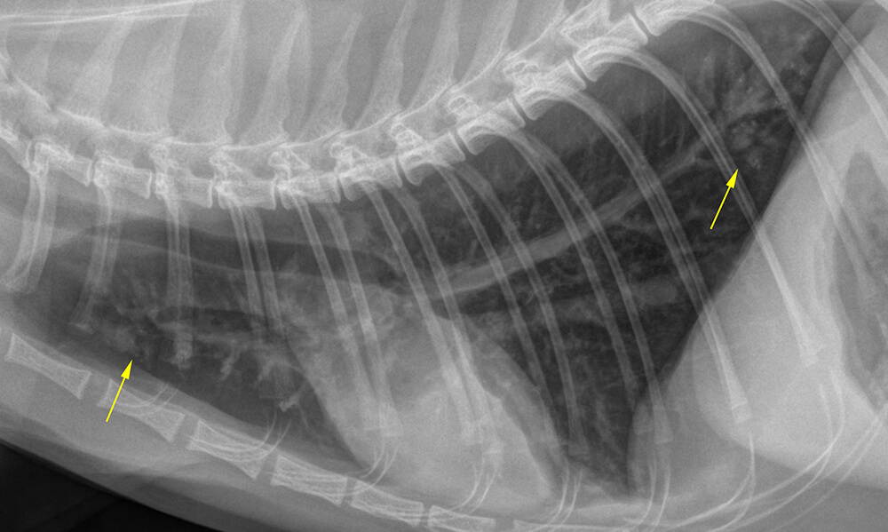
A 7-year-old DSH was presented for chronic intermittent cough which was only partially responsive to antimicrobial treatment. Radiographs revealed deep inflation of the lung in combination with a generalized bronchial and multinodular pattern.

CT findings
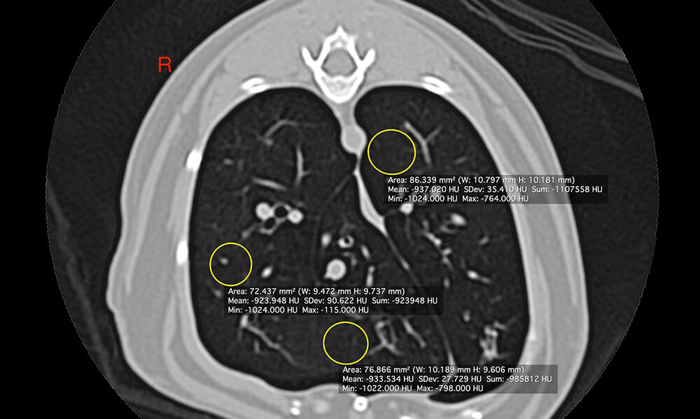
Expanded & overinflated lung with low attenuation values – average HU -930 – compatible with “Air-trapping”.
Moderate generalized bronchial pattern with multifocal cylindrical bronchiectasia in combination with mucus-plugging of branching bronchi and bronchioli resulting in a “Tree-in-bud sign”.
Lobar atelectasis of the caudal subsegment of the left cranial lung lobe with left-sided mediastinal shift.
Conclusions
The findings are typical for chronic feline lower airway disease. Allergic causes are most common. However, superinfection is a possibility. Bronchiectasia and atelectasis are commonly irreversible and may be associated with decreased mucociliary clearance rates which limits the potential for full recovery and increases the risk of superinfection.
Learning points
- The „Tree-in-bud sign“ reflects accumulation of fluid secretions within branching bronchi and/or bronchioli and is typical for chronic lower airway inflammation
- It may be mistaken for a nodular pattern on radiographs
- More information on the “Tree-in-bud sign” in cats is available in this recent article: Hahn et al. The computed tomographic “Tree‐in‐bud” pattern: Characterization and comparison with radiographic and clinical findings in 36 cats. Vet Radiol & Ultrasound 2017. https://onlinelibrary.wiley.com/doi/abs/10.1111/vru.12564
The diagnostic images – courtesy of Tierklinik Haar
UPLOAD MEDICAL IMAGES NOW